
ECG simulator
3D Electrocardiogram
Learn the ECG from the heart outward.
- Three languagesFull interface in English, Portuguese and Spanish
- Every screenRuns on desktop, tablet and smartphone
- Runs in your browser, nothing to installChrome 107+Edge 107+Safari 16+ · macOS, iPhone and iPadFirefox 104+An internet connection is required to load every application.
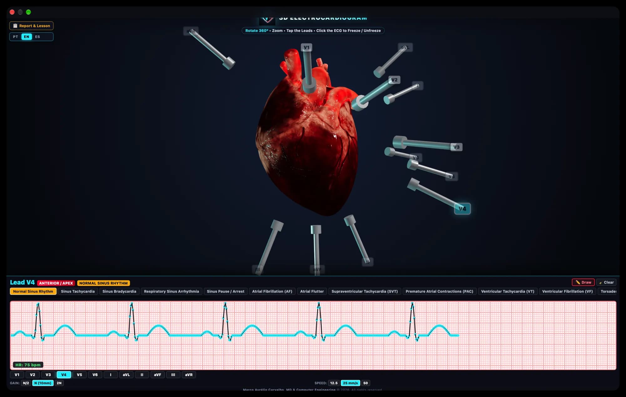
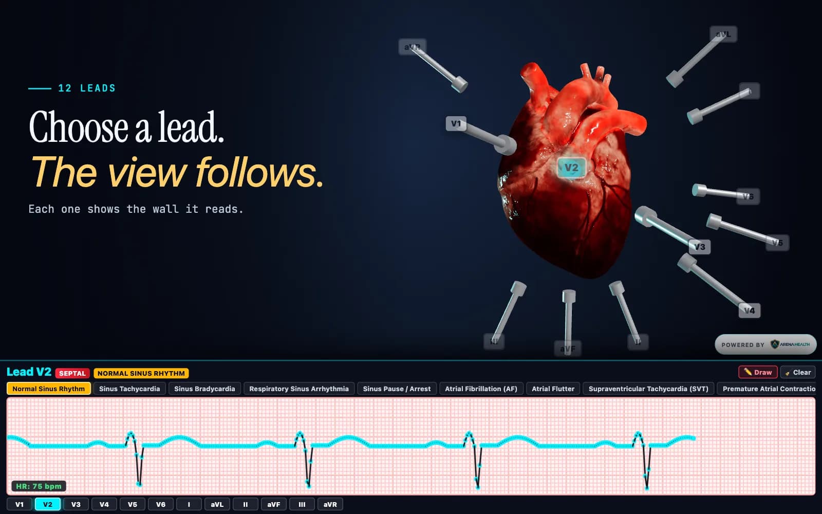
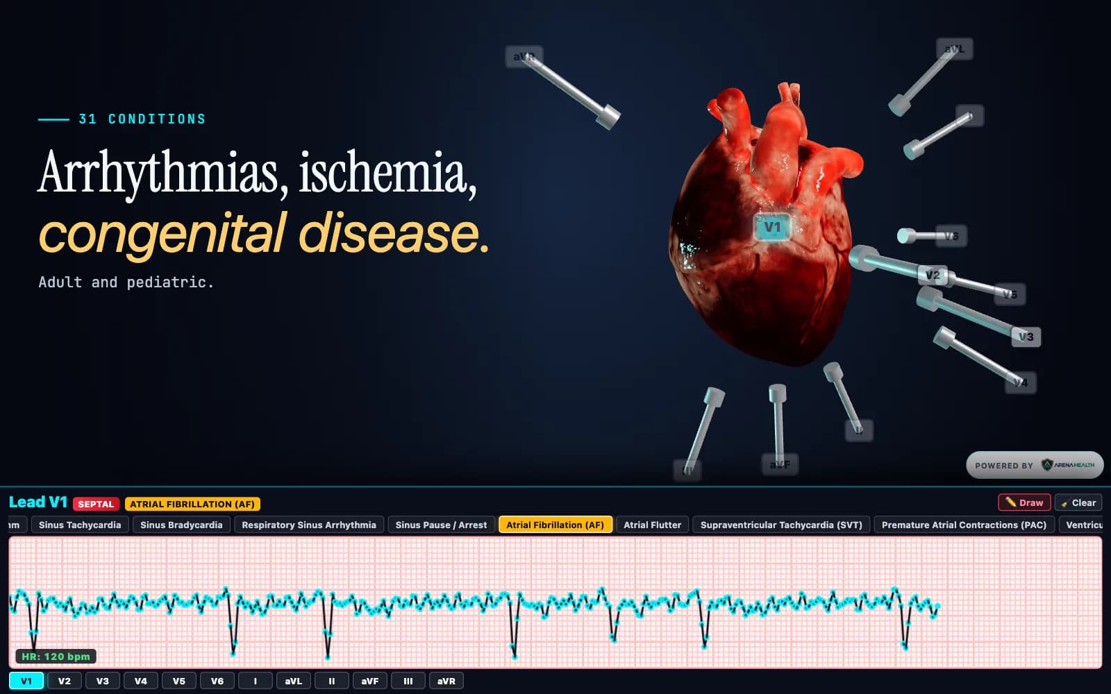
3D Electrocardiogram puts a beating 3D heart, its twelve electrodes and a live ECG monitor on one screen. Choose a lead and the camera moves to its electrode and names the wall it reads. Choose a rhythm and the trace changes, the heart beats in time with it, and a lesson explains what you are seeing in adults and in children.
- 12
- leads, each linked to its electrode and the wall it reads
- 31
- rhythms and conditions, adult and pediatric
- 7
- congenital heart defects, from ASD to hypoplastic left heart
Built by physicians.Grounded in science.Reviewed by senior clinicians.
Programmed, supervised and guided by physicians. Driven by computational physiology engines built from peer-reviewed research, and tested by senior healthcare professionals.
How we build our simulators3D Electrocardiogram on screen
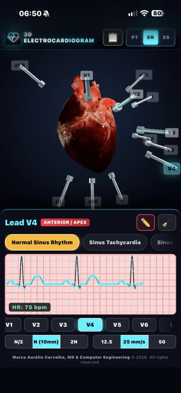
Phone layout 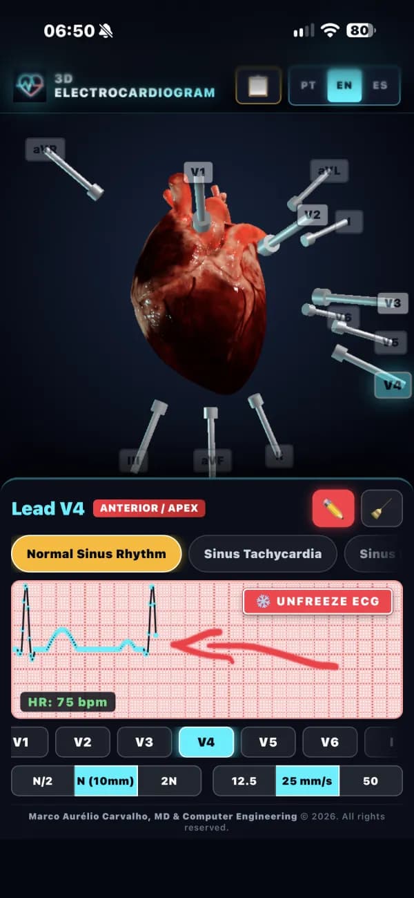
Phone layout
What it does
Everything below is in the app today.
A 3D heart with its leads
Electrodes and cables sit on a heart you can rotate through 360° and zoom. Choosing a lead moves the camera to its electrode.
Every lead mapped to a wall
V1–V6, I, II, III, aVR, aVL and aVF, each linked to the wall it explores: septal, anterior, apex, low and high lateral, inferior or the cavity.
Rhythms from sinus to arrest
Sinus tachycardia and bradycardia, respiratory sinus arrhythmia, sinus pause, atrial fibrillation and flutter, SVT, premature beats, VT, VF and torsades de pointes.
Conduction blocks
First-degree, Mobitz I with its Wenckebach pattern, Mobitz II, complete heart block, and left and right bundle branch block.
Infarction as it evolves
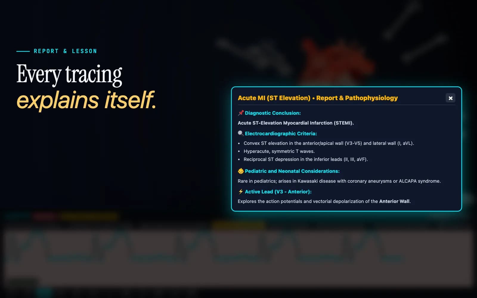
Acute STEMI with reciprocal changes, the subacute phase with Q waves and T inversion, and the chronic scar, alongside left and right ventricular hypertrophy.
Congenital heart disease
ASD, VSD, PDA, hypoplastic left heart, transposition of the great arteries, total anomalous pulmonary venous return and AV septal defect, each with its characteristic tracing.
A lesson behind every rhythm
A report for each rhythm gives the diagnostic conclusion, the criteria to look for (Sokolow-Lyon, Katz-Wachtel, rSR′, sawtooth flutter waves), the pathophysiology, and pediatric and neonatal considerations.
A monitor that behaves like one
Gain at N/2, N or 2N and paper speed at 12.5, 25 or 50 mm/s, with a live heart rate. Click to freeze the trace and draw on it to mark intervals and waves.
Bring your own heart model
Load your own .glb or .gltf heart in place of the built-in one.
An ECG class with 3D Electrocardiogram
On a projector or on each student’s phone, a rhythm takes a few minutes to teach.
- 1
Pick a rhythm
Choose from 31 conditions, from normal sinus rhythm to a complete AV septal defect.
- 2
Find the lead
Tap a lead to fly the camera to its electrode and see which wall of the heart it reads.
- 3
Freeze and mark
Stop the trace, change gain or speed, and draw on it to point out intervals and waves.
- 4
Open the lesson
Go through the criteria and the pathophysiology behind what the class just saw.
- 5
Compare adult and child
Use the pediatric and neonatal notes to show how the same finding reads in a younger patient.
Who it’s for and where it runs
Built for
- Medical students and residents learning ECG interpretation
- Pediatric, neonatal and cardiology teams
- Instructors teaching lead placement and rhythm recognition
Runs on
- Runs in the browser and installs to the home screen
- Phone layouts for portrait and landscape
- Respects reduced-motion settings
- English, Portuguese and Spanish
See 3D Electrocardiogram with your team.
Email us and we’ll arrange a walkthrough for your program, department or institution.