
Neuroimaging simulator
NeuroSim 3D
One brain, seen on CT, MRI and ultrasound.
- Three languagesFull interface in English, Portuguese and Spanish
- Every screenRuns on desktop, tablet and smartphone
- Runs in your browser, nothing to installChrome 107+Edge 107+Safari 16+ · macOS, iPhone and iPadFirefox 104+An internet connection is required to load every application.
Live in January 2027
Most neuroimaging teaching shows finished images, so students never see how a lesion becomes a signal. NeuroSim 3D starts from one shared 3D brain, places a pathology in it and computes each CT and MRI with its own physics engines. Change the sequence, the contrast or the day and the image is simulated again.
- 150
- conditions simulated in the pathology library
- 8
- MRI sequences and maps, at 1.5 T and 3 T
- 1 h–90 d
- timeline for staged hemorrhage and infarct
Built by physicians.Grounded in science.Reviewed by senior clinicians.
Programmed, supervised and guided by physicians. Driven by computational physiology engines built from peer-reviewed research, and tested by senior healthcare professionals.
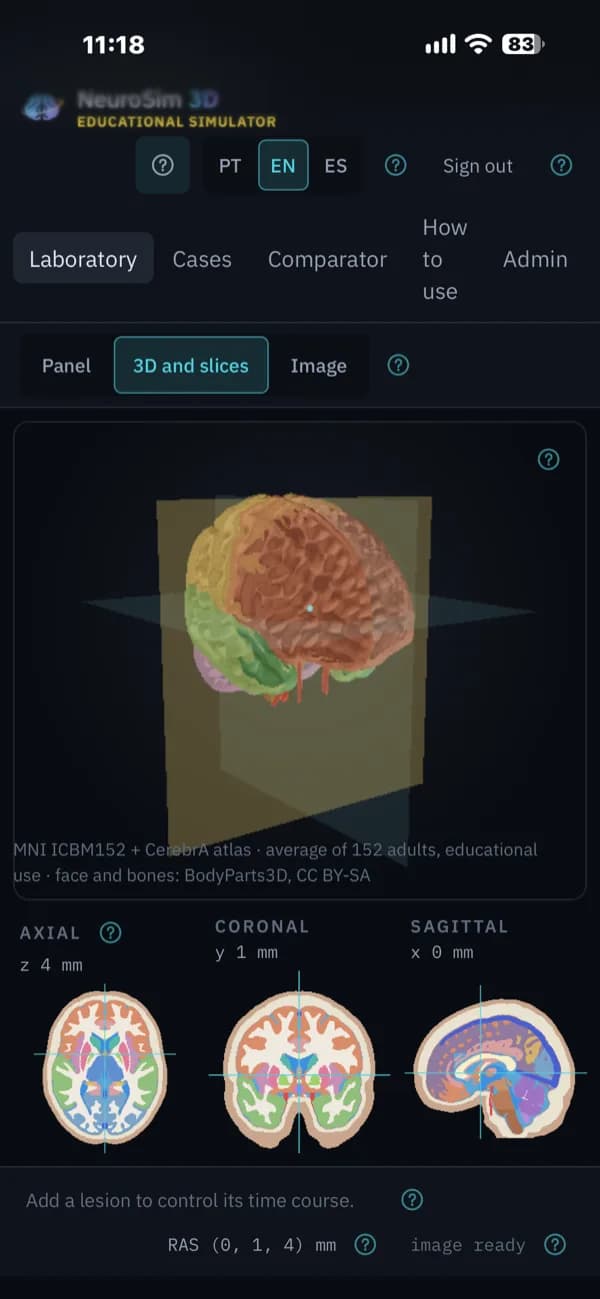
How we build our simulatorsNeuroSim 3D on screen
Phone layout 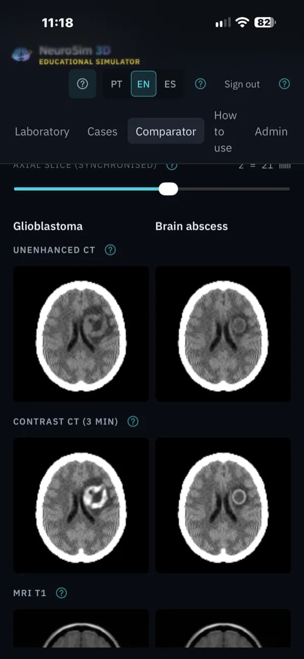
Phone layout 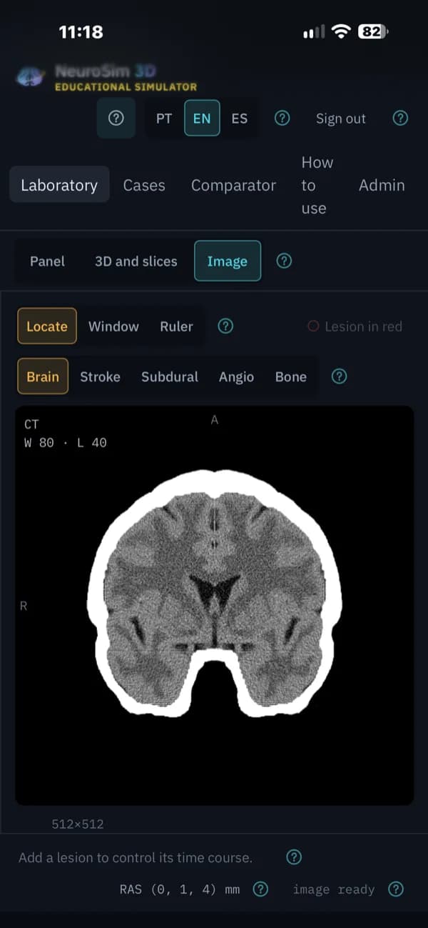
Phone layout
What it does
Everything below comes with the January 2027 launch.
A 3D brain you can take apart
Rotate, zoom and pan, change the opacity of each structure, hide or isolate it, explode the model or clip through it. Fade the cortex to reveal nuclei, limbic structures and ventricles.
Click to identify
Select a structure in the 3D model, on a slice or on a CT or MRI image to see what it is. Axial, coronal and sagittal slices stay in sync with the 3D view and the acquisition plane.
CT from projection physics
CT images are calculated by projecting through the anatomy and reconstructing with filtered back-projection, and can be read with windowing and Hounsfield values.
MRI across sequences
T1, fat-suppressed T1, T2, PD, FLAIR, T2*, DWI and ADC at 1.5 T or 3 T, all generated from the same anatomy, so the same slice can be compared in every contrast.
IV contrast and perfusion
Iodine on CT and gadolinium on MRI, as a single phase or a dynamic series: CT perfusion, multiphase CT, DSC and DCE, with time–intensity curves and extended Tofts kinetics. ASL is included.
A pathology library
Tumors, hemorrhage, infarct, abscess, hydrocephalus and extra-axial collections, built from 13 lesion mechanisms that alter the anatomy before any image is simulated.
Lesions over time
Move hemorrhage and infarct along a timeline from 1 hour to 90 days and every modality is simulated again for that moment.
Neonatal ultrasound
Transfontanellar imaging on preterm and term newborn anatomy, in coronal and sagittal planes, with control of angle, frequency, depth, focus, gain, TGC and dynamic range.
Compare and follow a guided case
A neuroradiology comparator sets images side by side, and a seven-step guided case walks through advanced contrast and perfusion.
Cases and grading
Instructors build a case from the lab and attach a question. Students run it and get feedback and a grade, while the answer key stays hidden.
A neuroimaging class with NeuroSim 3D
One case can carry a class from anatomy to diagnosis.
- 1
Start with the anatomy
Open the 3D brain and isolate the structures the lesion will affect.
- 2
Place the pathology
Choose a condition from the library for the class to study.
- 3
Acquire CT and MRI
Run the same slice through CT and each MRI sequence and compare what each one shows.
- 4
Inject contrast
See what enhances and read the time–intensity curves.
- 5
Move through time
Follow a hemorrhage or an infarct from the first hours to the following months.
- 6
Ask for the diagnosis
Assign a case with a question and review each student’s answer and grade.
Who it’s for and where it runs
Built for
- Radiology, neurology and neurosurgery residents
- Medical students learning neuroimaging
- Neonatal teams learning transfontanellar ultrasound
Runs on
- Runs in the browser, with nothing to install
- Desktop, tablet and phone layouts
- English, Portuguese and Spanish
See NeuroSim 3D with your team.
Email us and we’ll arrange a walkthrough for your program, department or institution.