

Ultrasound simulator
POCUS Sim 3D
Learn to scan before you touch a patient.
- Three languagesFull interface in English, Portuguese and Spanish
- Every screenRuns on desktop, tablet and smartphone
- Runs in your browser, nothing to installChrome 107+Edge 107+Safari 16+ · macOS, iPhone and iPadFirefox 104+An internet connection is required to load every application.
Live in January 2027
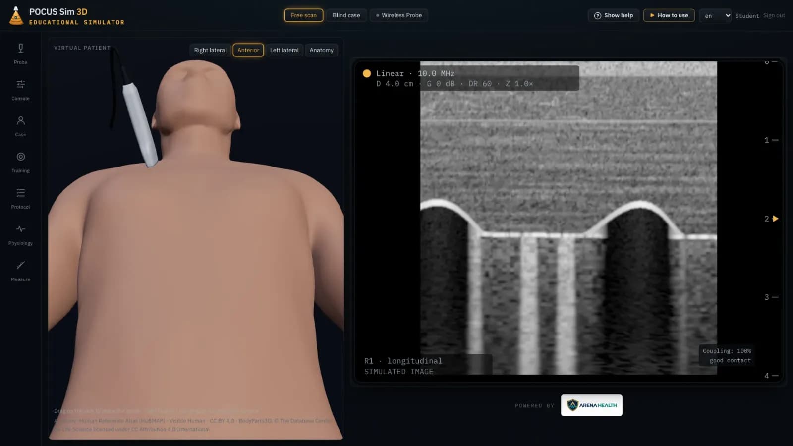
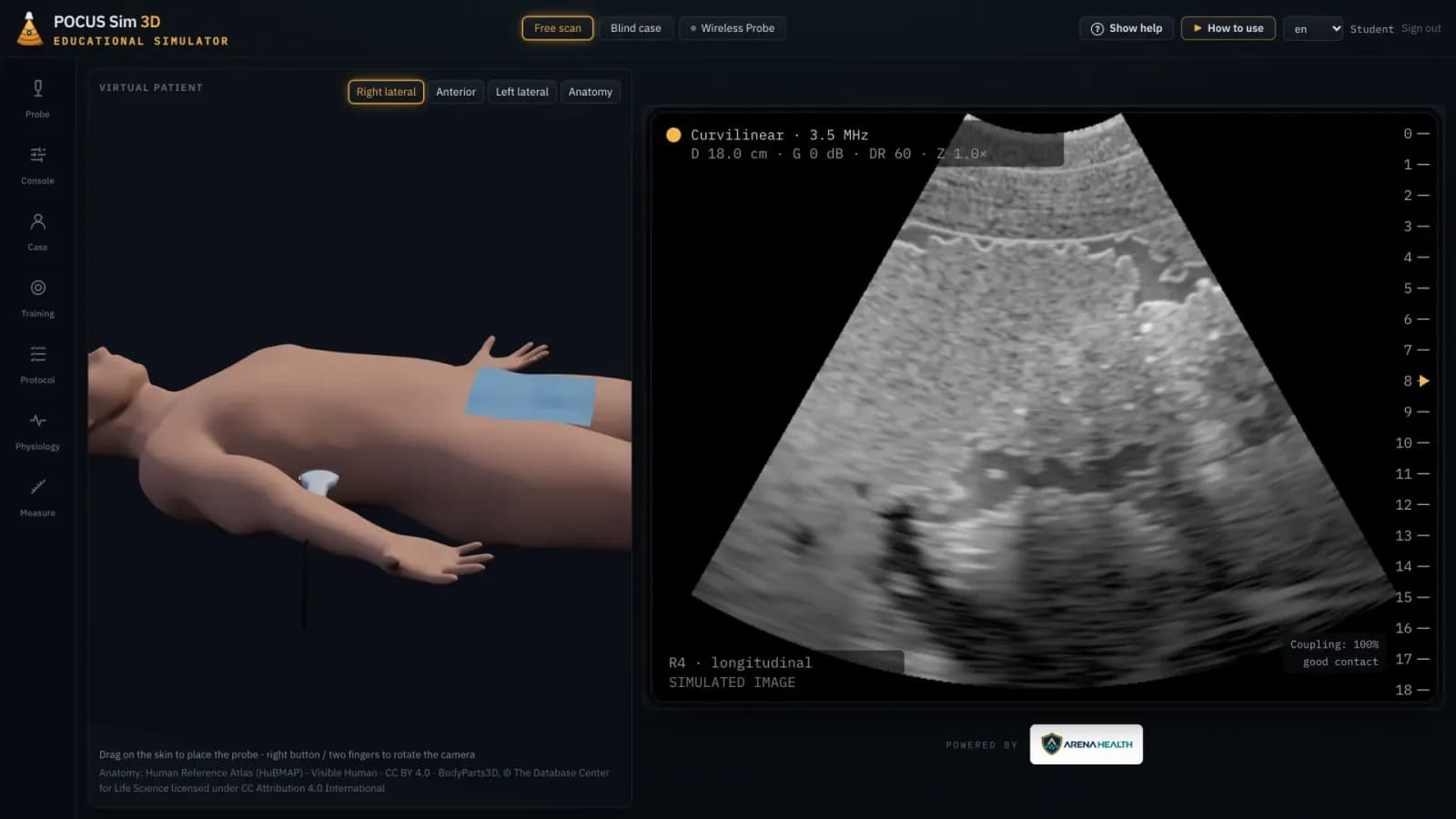
Most ultrasound teaching relies on recorded clips: you watch the image, but you never produce it. POCUS Sim 3D is a point-of-care ultrasound simulator in the browser. Move the probe over a 3D patient and the image is computed live from the anatomy under the beam.
- 9
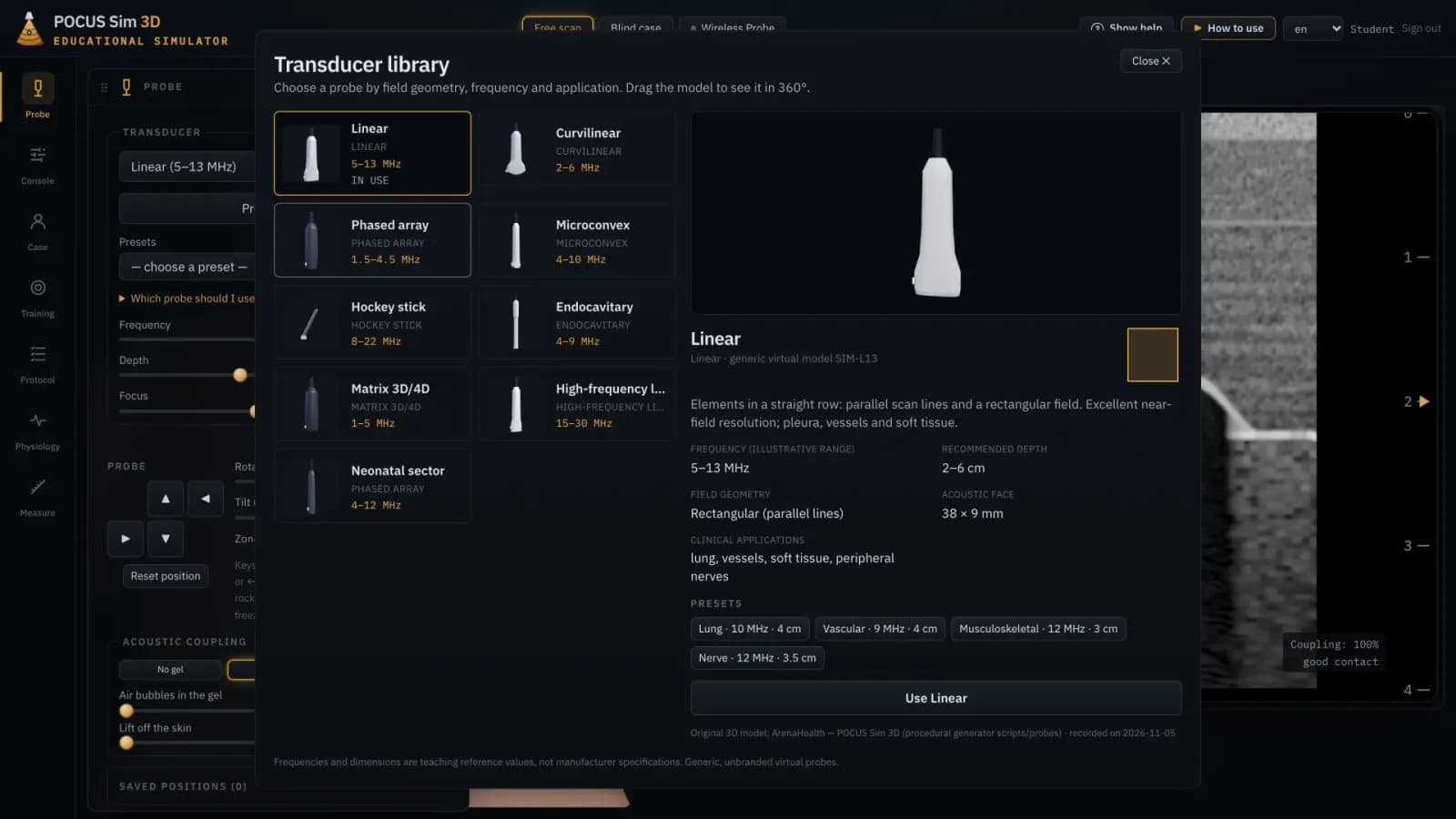
- virtual transducers, each with its own geometry
- 11
- echocardiographic views on a beating heart
- 5
- Doppler modes, from color to tissue
Built by physicians.Grounded in science.Reviewed by senior clinicians.
Programmed, supervised and guided by physicians. Driven by computational physiology engines built from peer-reviewed research, and tested by senior healthcare professionals.
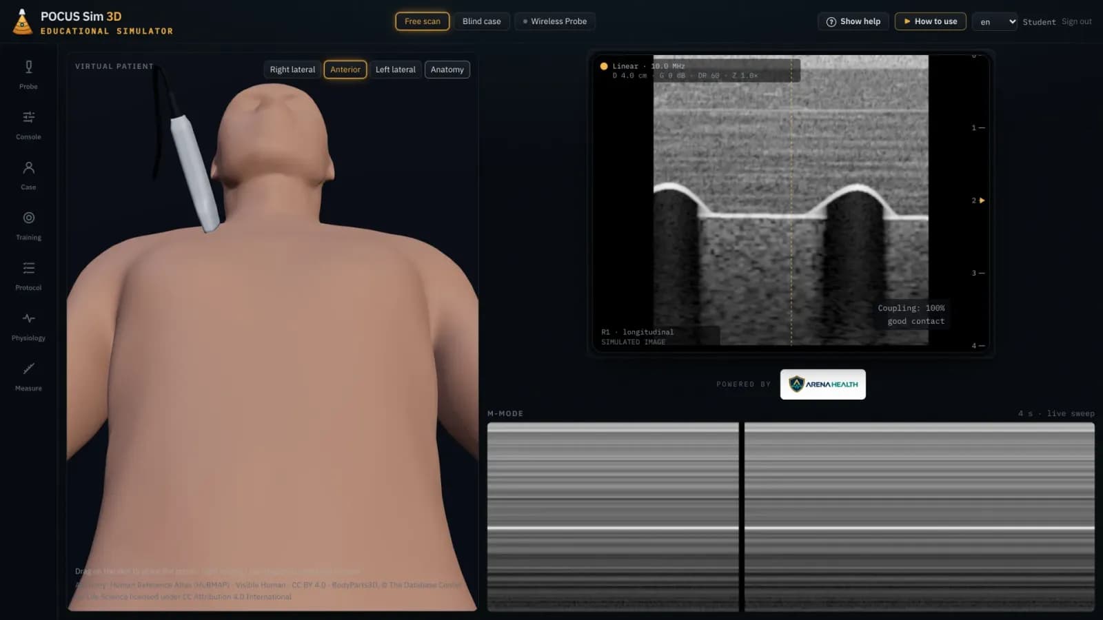
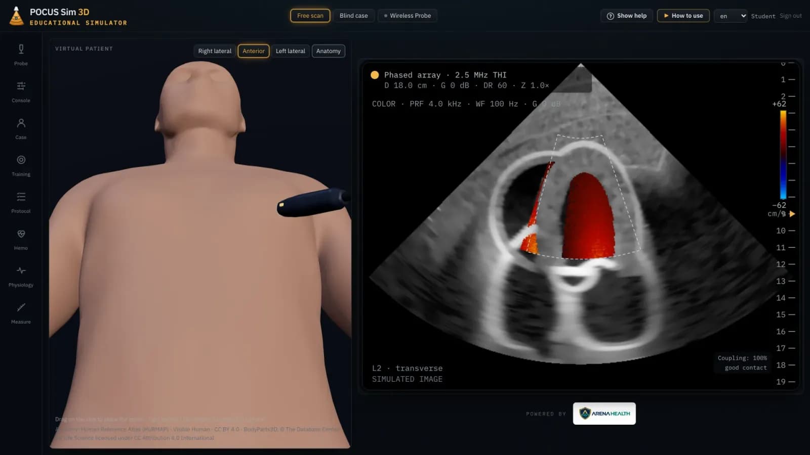
How we build our simulatorsPOCUS Sim 3D on screen
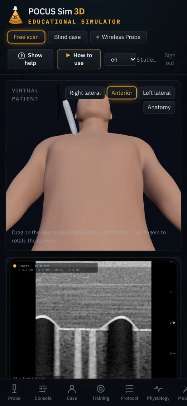
Phone layout
What it does
Everything below comes with the January 2027 launch.
Not a video library
Slide, rotate, tilt, rock or press and the image responds. Rib shadows, A-lines and B-lines appear because of where the probe is, not because a clip was played.
Lung
Work through the chest zone by zone. Recognize A-lines and B-lines, pleural effusion and consolidation, and use M-mode to study pneumothorax.
Heart
Acquire 11 echocardiographic views on a beating heart, including parasternal long and short axis, apical, subcostal and suprasternal windows.
Doppler
Switch between color, power, pulsed-wave, continuous-wave and tissue Doppler, and read the spectral trace as it builds.
Abdomen and FAST
Scan the right upper quadrant and other abdominal windows, and look for free fluid in trauma scenarios.
Advanced hemodynamics
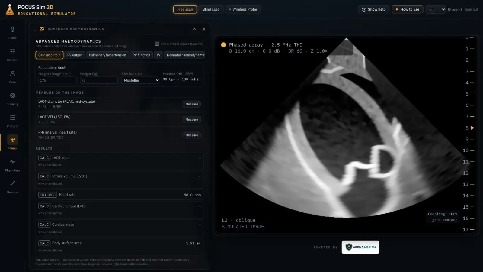
Take measurements on the simulated image and carry them through to stroke volume, cardiac output and right-heart assessment. Each result shows whether it was measured, entered, calculated or estimated.
The anatomy behind the image
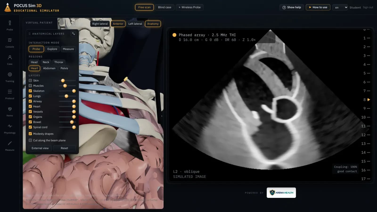
Peel away skin and muscle, isolate the heart or lungs, and cut the 3D model along the beam to see exactly which structures the image passes through.
The right probe
Nine virtual transducers, from linear and curvilinear to phased array and neonatal sector, each with its own field geometry, frequency range and clinical applications.
From free practice to assessment
Scan freely, follow FoCUS and RUSH step by step, find a target window for a score based on probe position, or scan an unknown patient, commit to a diagnosis and get feedback.
Measure, annotate, save
Measurements, annotations and image saving, on adult and neonatal patients.
A scanning session with POCUS Sim 3D
From free practice to a blind case, at the learner’s own pace.
- 1
Free scan
Pick a scenario and a probe and explore at your own pace.
- 2
Guided protocols
Follow FoCUS and RUSH step by step and record your findings.
- 3
Training exercises
Find the target window with the right probe and get a score based on your probe position.
- 4
Blind cases
Scan an unknown patient, commit to a diagnosis and get feedback.
Who it’s for and where it runs
Built for
- Medical students and residents learning point-of-care ultrasound
- Emergency, critical-care and neonatal teams
- Instructors running ultrasound courses
Runs on
- Runs in the browser, with nothing to install
- Works on desktop, tablet and phone
- English, Portuguese and Spanish
See POCUS Sim 3D with your team.
Email us and we’ll arrange a walkthrough for your program, department or institution.