

Veterinary neuroimaging simulator
NeuroSim VET
Many species, one imaging engine.
- Three languagesFull interface in English, Portuguese and Spanish
- Every screenRuns on desktop, tablet and smartphone
- Runs in your browser, nothing to installChrome 107+Edge 107+Safari 16+ · macOS, iPhone and iPadFirefox 104+An internet connection is required to load every application.
Live in January 2027
A veterinary student may see few CT or MRI studies of the brain, and fewer still in a horse or a pig. NeuroSim VET brings the NeuroSim physics engines to dog, cat, horse, cattle, sheep and pig models, with veterinary planes and names, so the same lesion can be studied across sequences and species.
- 6
- species, with seven brain models
- 65
- veterinary conditions with a working simulation
- 8
- MRI sequences and maps, at 1.5 T and 3 T
Built by physicians.Grounded in science.Reviewed by senior clinicians.
Programmed, supervised and guided by physicians. Driven by computational physiology engines built from peer-reviewed research, and tested by senior healthcare professionals.
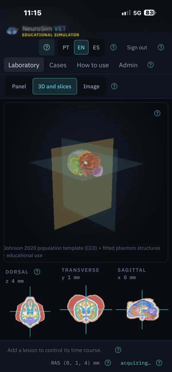
How we build our simulatorsNeuroSim VET on screen
Phone layout 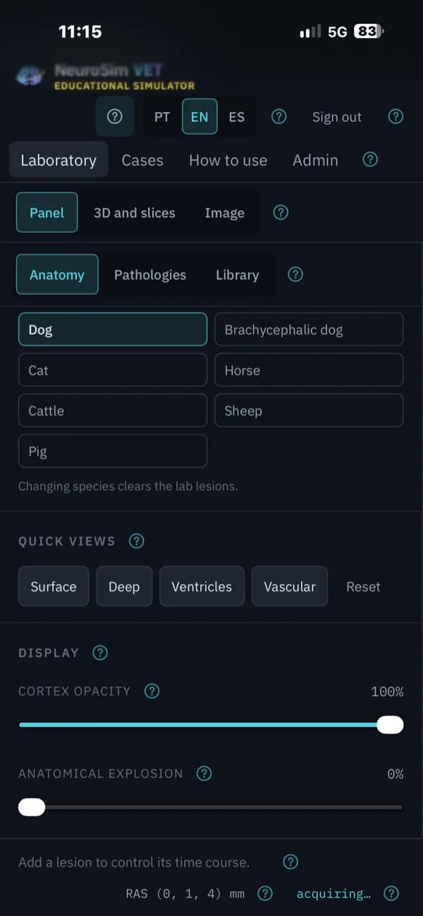
Phone layout 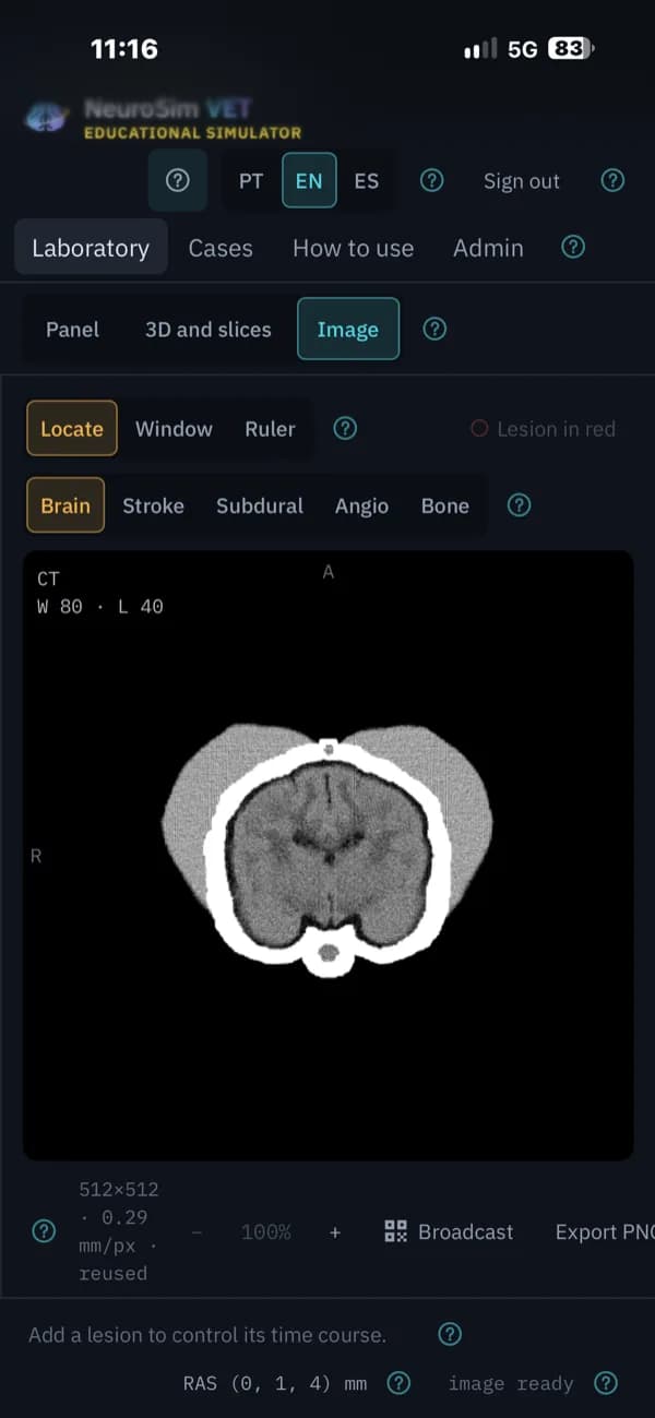
Phone layout
What it does
Everything below comes with the January 2027 launch.
Seven brains, six species
Mesaticephalic and brachycephalic dogs, cat, horse, cattle, sheep and pig, each with its own head proportions, such as the bony tentorium of the cat or the pneumatized frontal sinus of cattle.
Veterinary planes and names
Images are read as dorsal, transverse and sagittal, and structures use veterinary terms such as dorsal sagittal sinus and rostral and caudal cerebral arteries.
Look inside the brain
Fade the cortex in 3D to reveal nuclei, limbic structures and ventricles, on the anatomy of the species you are teaching.
CT and MRI from physics
The same CT and MRI engines as NeuroSim 3D, with sequences including T1, T2, FLAIR and DWI at 1.5 T or 3 T.
A veterinary pathology library
Conditions such as feline meningioma and congenital hydrocephalus, simulated across CT and MRI, with gadolinium contrast.
Canine ultrasound
Scan through a persistent bregmatic fontanelle, as in puppies and toy breeds, and find a congenital hydrocephalus or a quadrigeminal cyst.
A veterinary imaging class with NeuroSim VET
Compare species and sequences without waiting for the right case to arrive.
- 1
Pick the species
Open a dog, a cat or a large-animal model and explore it in 3D.
- 2
Choose a condition
Select a lesion from the veterinary library.
- 3
Acquire the images
Run CT and MRI in veterinary planes and compare the sequences.
- 4
Add contrast or ultrasound
Give gadolinium to see what enhances, or scan a dog through the fontanelle.
Who it’s for and where it runs
Built for
- Veterinary students learning neuroimaging
- Residents in veterinary neurology and diagnostic imaging
- Instructors teaching small- and large-animal cases
Runs on
- Runs in the browser, with nothing to install
- Desktop, tablet and phone layouts
- English, Portuguese and Spanish
See NeuroSim VET with your team.
Email us and we’ll arrange a walkthrough for your program, department or institution.